Foot Anatomy Basics

  • 01. The Plantar Fascia

    The Plantar Fascia

    The plantar fascia is a thick, fibrous band of connective tissue located along the bottom of the foot. It extends from the calcaneus (heel bone) to the base of the toes, forming a crucial part of the foot’s structure.

    Working alongside other ligaments and tendons, the plantar fascia helps support the foot’s arches and plays a vital role in absorbing shock during activities like walking, running, or jumping.

    It also helps maintain proper foot mechanics by stabilizing the foot during movement and distributing pressure evenly across the sole.

  • 02. Metatarsals

    Metatarsals

    Metatarsals are the five long bones located in the midfoot. They connect the phalanges to the tarsal bones near the ankle, forming the bridge between the forefoot and hindfoot.

    Each metatarsal is numbered one through five, starting with the first metatarsal, which is the thickest and shortest and connects to the big toe.

    These bones play a vital role in weight-bearing, balance, and walking, as they help transfer force from the heel to the toes during motion.

  • 03. Phalanges

    Phalanges

    Each foot contains a total of 14 phalanges. Phalanges are the bones that make up the toes of the foot, and they share the same anatomical name as the bones in the fingers of the hand. The big toe (also known as the hallux) has two phalanges, a proximal and a distal phalanx, while each of the other four toes has three phalanges: proximal, middle, and distal. These bones play a critical role in balance, walking, and supporting body weight during movement.

  • 04. Tarsals

    Tarsals

    Tarsal bones are a group of seven irregularly shaped bones located in the rearfoot and midfoot. These bones form the back portion of the foot and connect the foot to the lower leg, playing a crucial role in stability, mobility, and shock absorption.

    The largest of these bones is the calcaneus, commonly known as the heel bone, which bears most of the body's weight when standing or walking. Above the calcaneus sits the talus, which forms the lower part of the ankle joint and connects the foot to the tibia and fibula of the lower leg.

    The remaining five tarsal bones—the navicular, cuboid, and the three cuneiforms (medial, intermediate, and lateral)—fit together in a complex arrangement that allows the foot to be both strong and flexible. These bones form the arches of the foot, working together with the metatarsals to distribute weight, absorb impact, and provide leverage during movement.

  • 05. Arch Support

    Arch Support

    In addition to supporting body weight and aiding in movement, the metatarsals also contribute to the formation and maintenance of the foot’s natural arches, particularly the transverse and longitudinal arches. These arches are essential for shock absorption, balance, and efficient energy transfer with each step. Proper metatarsal alignment and function are critical for healthy arch support, helping prevent foot fatigue, discomfort, and injuries.

The Plantar Fascia

The plantar fascia is a thick, fibrous band of connective tissue located along the bottom of the foot. It extends from the calcaneus (heel bone) to the base of the toes, forming a crucial part of the foot’s structure.

Working alongside other ligaments and tendons, the plantar fascia helps support the foot’s arches and plays a vital role in absorbing shock during activities like walking, running, or jumping.

It also helps maintain proper foot mechanics by stabilizing the foot during movement and distributing pressure evenly across the sole.

Metatarsals

Metatarsals are the five long bones located in the midfoot. They connect the phalanges to the tarsal bones near the ankle, forming the bridge between the forefoot and hindfoot.

Each metatarsal is numbered one through five, starting with the first metatarsal, which is the thickest and shortest and connects to the big toe.

These bones play a vital role in weight-bearing, balance, and walking, as they help transfer force from the heel to the toes during motion.

Phalanges

Each foot contains a total of 14 phalanges. Phalanges are the bones that make up the toes of the foot, and they share the same anatomical name as the bones in the fingers of the hand. The big toe (also known as the hallux) has two phalanges, a proximal and a distal phalanx, while each of the other four toes has three phalanges: proximal, middle, and distal. These bones play a critical role in balance, walking, and supporting body weight during movement.

Tarsals

Tarsal bones are a group of seven irregularly shaped bones located in the rearfoot and midfoot. These bones form the back portion of the foot and connect the foot to the lower leg, playing a crucial role in stability, mobility, and shock absorption.

The largest of these bones is the calcaneus, commonly known as the heel bone, which bears most of the body's weight when standing or walking. Above the calcaneus sits the talus, which forms the lower part of the ankle joint and connects the foot to the tibia and fibula of the lower leg.

The remaining five tarsal bones—the navicular, cuboid, and the three cuneiforms (medial, intermediate, and lateral)—fit together in a complex arrangement that allows the foot to be both strong and flexible. These bones form the arches of the foot, working together with the metatarsals to distribute weight, absorb impact, and provide leverage during movement.

Arch Support

In addition to supporting body weight and aiding in movement, the metatarsals also contribute to the formation and maintenance of the foot’s natural arches, particularly the transverse and longitudinal arches. These arches are essential for shock absorption, balance, and efficient energy transfer with each step. Proper metatarsal alignment and function are critical for healthy arch support, helping prevent foot fatigue, discomfort, and injuries.

Understandıng MOVEMENT

01 Ankle

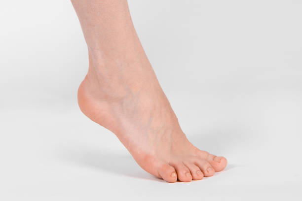
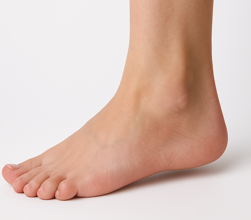
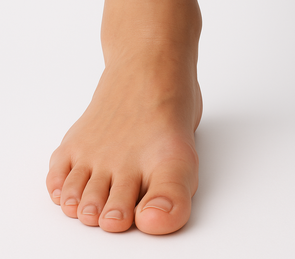
It moves in two directions. Plantar flexion is when you’re sitting with your legs out infront of you and you point your toes forward and down. And Dorsiflexion is when you unpoint your toes and your feet come back to normal position with toes pointing upwards. It’s basically your natural standing position. 

01 Ankle

02 Middle Foot

The metatarsal area allows for two movements which are supination and pronation. Or they can also be called inversion or eversion. This is when the weight of your body shifts from side to side and your feet hold the weight by turning in or out.

02 Middle Foot

03 Toes

The toes can do four moves. They flex (bend) when you scrunch a towel or pick something up with your toes. They extend (straighten) this is when you go onto your tip toes. They abduct which is when you splay your toes out and they lastly adduct which is when you squeeze them together. Or when they’re unhappily squeezed into a narrow shoe!

03 Toes

Benefits of stronger feet

Unfortunately, we don’t use our feet as much as we do our hands and fingers. And as a result, the strength in our toes becomes limited and as we get older, individual toe movements become weak and unable to do what it’s supposed to do. It’s like all the muscles become stagnant and stuck together which causes inflammation and pain.

Strong feet and toes lead to more muscle movement and stronger upward joints. Plus, deterity in toes is important because it supports better balance and stabilizes the entire body.

Barefoot shoes

Wearing barefoot shoes that have a wide toe box and flexible sole is key to maintaining natural foot alignment. Wearing narrow shoes pushes the big toe inwards which counteracts what the toe spacer is meant to do. You can check out Nunorm barefoot shoes here.